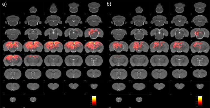
Fig 1. Brain regions with significant connections with the bilateral RSCs in the anesthetized rats: (a) left RSC and (b) right RSC.
These significant regions were shown in coronal slices as a color-coded statistical T-values superimposed on a set of normalized coronal atlas of the rat brain. RSC, the retrosplenial cortex.

