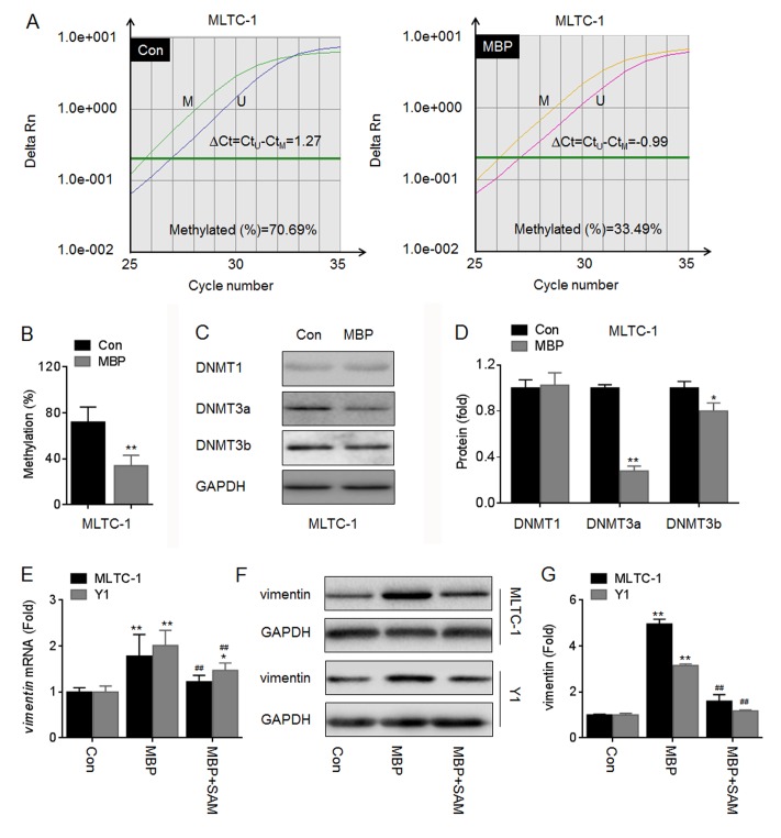
Fig 6. MBP improved the expression of vimentin by DNA demethylation.
(A-D), MLTC-1 cells were exposed to 1000 nM MBP for 24 h, the methylation status of vimentin promoter was determined in triplicate by qMSP (A and B). Annotation, methylated (M); unmethylated (U); the percentage of methylation in a sample was estimated using the following formula: methylation (%) = (M/M+U) ×100% = [1/(1+U/M)] ×100% = [1/(1+2∆Ct)] ×100%. (C and D), Western blots analysis and relative protein levels of DNMT1, DNMT3a, and DNMT3b. *p<0.05 and **p<0.01 compared with medium control cells. (E-G), MLTC-1 and Y1 cells were exposed to 1000 nM MBP in the presence or absence of 200 μM SAM for 24 h, respectively. (E) qRT-PCR analysis in triplicate of vimentin mRNA. (F) Western blots analysis and (G)relative protein levels of vimentin. *p<0.05 and **p<0.01 compared with medium control cells; ##p<0.01 compared with cells exposed to MBP alone.

