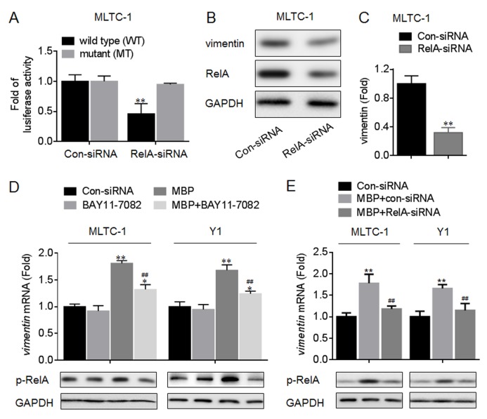
Fig 8. Functions of NF-κB in the transcriptional activation of vimentin.
(A) MLTC-1 cells were co-transfected by Con-siRNA or RelA-siRNA plus pGL3-vimentin-Luc construct (wild type, WT; or mutant, MT) for 12 h. Luciferase reporter assay analysis of the effects of NF-κB on the transcriptional activity in vimentin promoter. (B and C) MLTC-1 cells were transfected by Con-siRNA or RelA-siRNA for 12 h. (B) Western blots analysis and (C)relative protein levels of vimentin. **p<0.01 compared with cells transfected by Con-siRNA. (D) After MLTC-1 and Y1 cells were pre-treated by 0 or 10 μM BAY11-7082 for 12 h, they were exposed to 0 or 1000 nM MBP for 24 h. (D, top) qRT-PCR analysis in triplicate of vimentin mRNA, *p<0.05 and **p<0.01 compared with medium control cells; ##p<0.01 compared with cells exposed to MBP alone. (D, bottom) Western blots analysis of the expression of p-RelA. (E) After MLTC-1 and Y1 cells were pre-transfected by Con-siRNA or RelA-siRNA for 12 h, they were exposed to 1000 nM MBP for 24 h. (E, top) qRT-PCR analysis in triplicate of vimentin mRNA, **p<0.01 compared with medium control cells; ##p<0.01 compared with cells exposed to MBP plus Con-siRNA.

