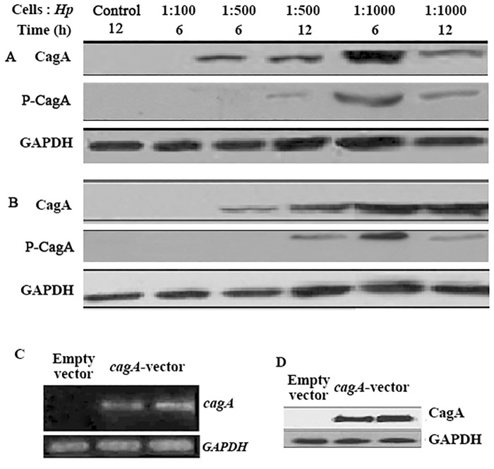
Fig 1. Introduction of CagA into gastric cancer cells.
(A and B) Western blot analysis of CagA and phosphorylated CagA in H. pylori-infected SGC-7901(A) and AGS (B) cells. The cells infected with the indicated ratio of cells to H. pylori for the indicated time were collected and lysed, and the proteins were separated by SDS-PAGE. Cells infected with H. pylori boiled for 15 min at a MOI of 1:1000 were used as a control. (C and D) Detection of CagA mRNA and protein in cagA-overexpressing SGC-7901 cells by RT-PCR (C) and western blot (D). GAPDH served as the loading control. The data are representative of three independent experiments. Hp, H. pylori; P-CagA, phosphorylated CagA; GAPDH, Glyceraldehyde-3-phosphate- dehydrogenase.

