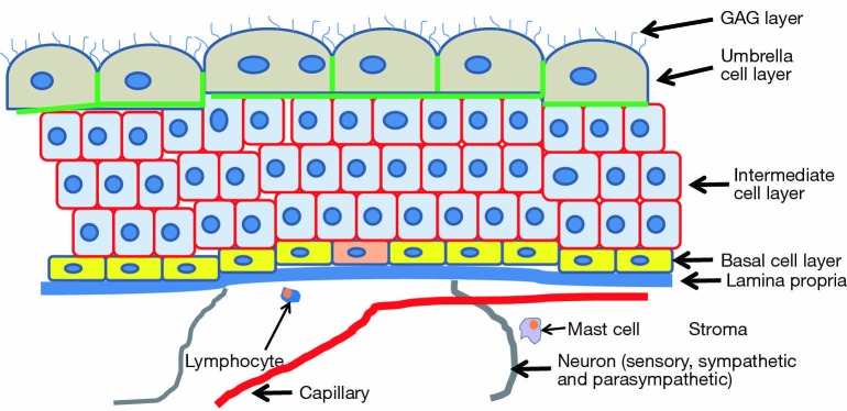
Figure 1.
Schematic illustration of bladder anatomy. The stroma is composed of the detrusor muscle, connective tissue, nerves, and the veins, arteries and capillaries of the connective system. The urothelium sits atop the lamina propria. The nerves, and capillaries do not penetrate the lamina propria but do connect with it. Therefore the bladder urothelium is less well supplied than are other epithelia. Also found are neurons, including the sensory, sympathetic and parasympathetic systems. These generally are found adjacent to the lamina propria, indicating the lamina may respond to neural signals. The basal layer is the lowest layer of cells. These are generally the only cells that divide. The stem cells, here one is shown in pink, also exist within the basal cell layer. Atop the basal cells is a layer of 3-5 cells deep comprised of intermediate cells. Not shown is that these cells maintain a connection via a cytoplasmic process to the lamina propria (not shown). Most likely this connection serves a cell communication function. The outer layer of cells, or umbrella cells, are highly specialized and terminally differentiated. They may be multinucleated as the result of cell fusion. Their lifetime is as much as 6 months or more. Loss of an umbrella cell immediately leads to the underlying intermediate cell to differentiate and replace the lost umbrella cell. The umbrella cells are coated with a dense layer of GAG, mostly if not exclusively, chondroitin sulfate. They also have tight junctions here shown in red along their basolateral surfaces. Together the uroplakin plaques (not shown) and GAG on the apical surface plus the tight junctions combine to make the urothelium the least permeable mammalian epithelium. GAG, glycosaminoglycan.

