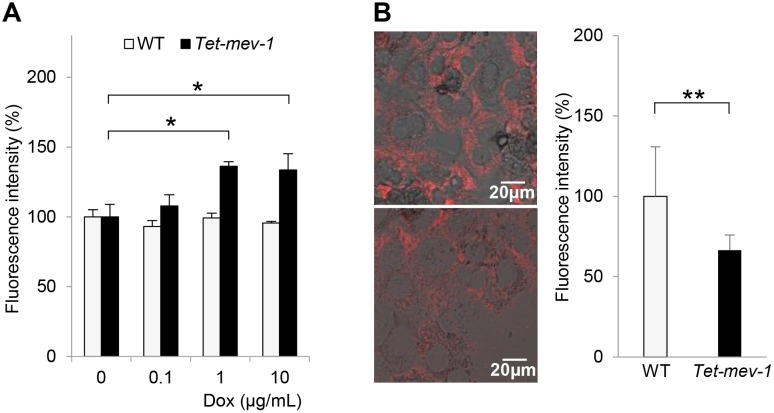
Fig 3. Increased reactive oxygen species (ROS) production and decreased mitochondrial membrane potential in Tet-mev-1 hepatocytes.
(A) Primary hepatocytes were isolated from 14-month-old male wild type (WT) or Tet-mev-1 mice that had been supplied with doxycycline-free water. After treatment with the indicated concentrations of doxycycline (Dox) for 72 hours, they were incubated with 100 μM dichlorofluorescein diacetate for 30 min. The fluorescence intensity was measured at 520 nm, and the values are expressed as means ± SD from six samples in each group. (B) Hepatocytes that had been isolated from the same mice used for the experiment shown in Fig 3A were incubated with 300 nM tetramethyl rhodamine methyl ester for 30 min. The fluorescence at 573 nm was viewed and analyzed using a confocal laser-scanning microscope with the same excitation strength and detection gain. Representative images are shown for hepatocytes obtained from wild type (upper) and Tet-mev-1 mice (lower). Scale bars, 20 μm. On the right side of the images, histograms indicating the values (means ± SD) of fluorescence intensities obtained from five randomly selected visual fields in each group are shown. The asterisks indicate that the differences between the groups are statistically significant (*p < 0.01; **p < 0.05).

