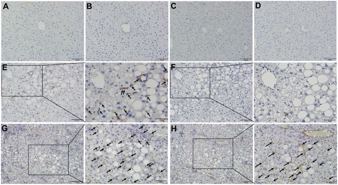
Fig 10. Increased infiltration of CCR5-positive cells and activation of hepatic stellate cells in liver tissues of Tet-mev-1 mice.
Liver specimens were obtained from male wild type (A, B, E and F) or Tet-mev-1 mice (C, D, G and H) that had been supplied with doxycycline-containing water and were subsequently fed control, normal chow (A to D) or a high-fat/high-sucrose diet (E to H) for 4 months at around the age of 2 years. The tissue sections were subjected to immunohistochemical staining using antibodies against CCR5 (A, C, E and G) or α-smooth muscle actin (B, D, F and H). CCR5-positive cells are shown by arrows in panels E and G, while α-smooth actin-expressing myofibroblasts were indicated by arrows in panel H. Representative images are shown from five mice in each group. Scale bars, 100 μm. A part of panels E to H is presented under high magnification in the corresponding panels on the right.

