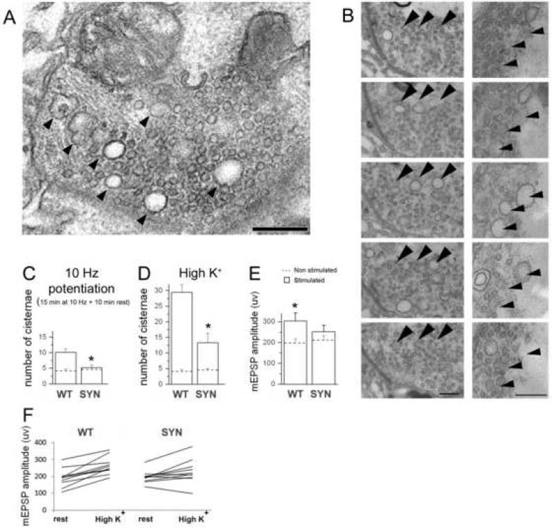
Figure 8. Synapsin promotes a formation of enlarged vesicles and quanta.
A. Endosome-like structures (arrowheads) formed upon sustained depolarization.
B. Tracing several endosome-like structures (arrowheads) through subsequent serial sections demonstrates that they are not connected to the plasma membrane or mitochondria.
B. Potentiation activates formation of endosome-like structures/enlarged vesicles in WT but not in synapsin (−) boutons. Asterisk indicate significant difference (p<0.05) between WT and synapsin (−) genotypes upon potentiation. Dotted lines – before stimulation; bars – after stimulation.
C. High K+ treatment drastically increases the number of endosome-like structures/enlarged vesicles in WT boutons, and this effect is not as prominent in synapsin (−) boutons. EM data collected from 3 preparations (16-19 boutons) for each line at each condition.
D, F. After high K+ treatment, quantal size significantly increases in synapsin (+) but not in synapsin (−) boutons, as could be seen from mean mEPSP amplitudes (D) or those in individual experiments (F). Asterisks indicate significant difference (p<0.05) between WT and synapsin (−) high K+ treated preparations. Data collected from 22 WT and 26 synapsin (−) preparations.

