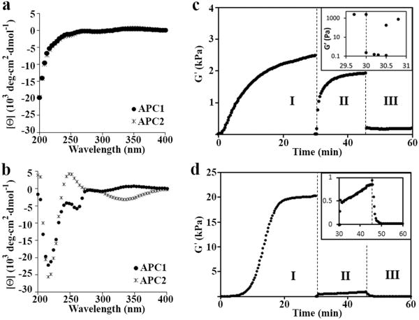
Figure 3. Biophysical and mechanical characterization of the hydrogel.
(a) CD wavelength spectra of 1 wt% APC1 and APC2 in water at 5 °C showing that the peptides are unfolded. (b) Wavelength spectra of 1 wt% APC1 and APC2 hydrogels formed at pH 7.4 (150 mM NaCl) and 25 and 37 °C, respectively, indicating peptide folding and assembly. (c) Rheological assessment showing the formation, shear-thin/recovery and photodisruption of APC1 and (d) APC2 gels. In regime I, a dynamic time sweep monitors the initial sol-to-gel phase transition of each 1 wt% gel after folding and assembly is triggered by measuring the storage modulus (G′) as a function of time at pH 7.4 and 37 °C for APC1 and 25 °C for APC2; frequency = 6 rad/s, 0.2% strain. In regime II, 1000% strain is applied for 30 seconds to thin the materials after which the strain is decreased to 0.2% to allow material recovery. Inset in panel (c) shows regimes I and II expanded, highlighting the gel-sol and subsequent sol-gel transitions. In regime III, the recovered gels are irradiated at 365 nm for 10 minutes to initiate the final gel-sol transition; G′ is monitored during and after irradiation. Inset in panel (d) shows regimes II and III expanded. Independent frequency- and strain-sweep measurements (Figures S5 and S6), additional rheology experiments on pre-formed gels (Figure S7), and AFM measurements (Table S1) further support this rheological profile.

