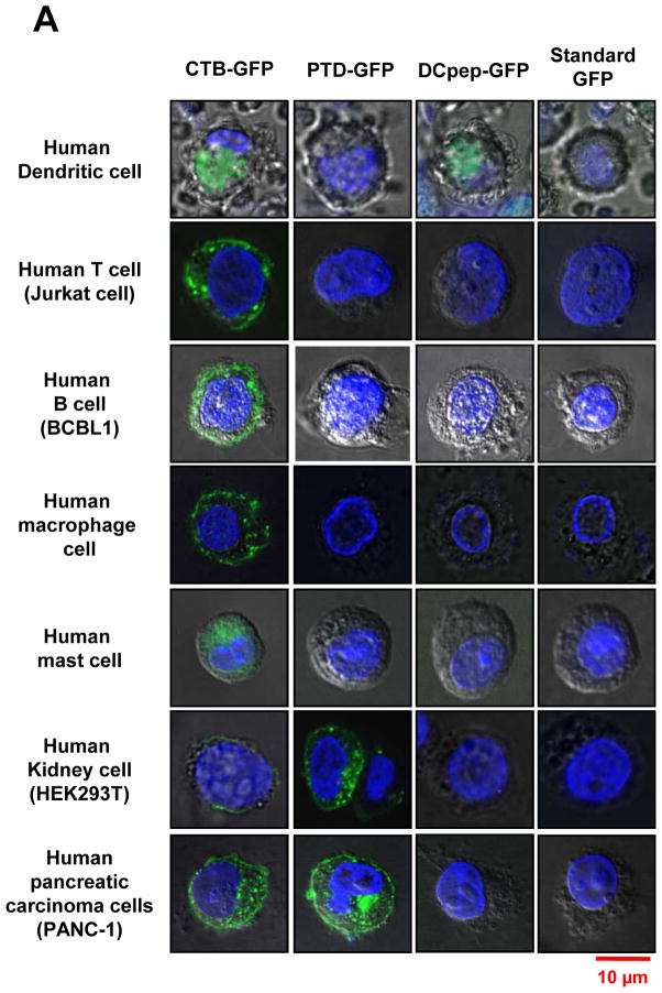
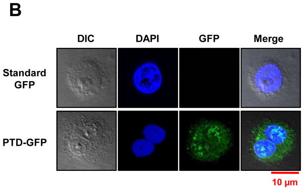
Fig. 5. Uptake of GFP fused with different tags by human immune and non-immune cells.
(A) Translocation of purified GFP fusion proteins in human cell lines. 2×10⁴ cells of cultured human dendritic cell (DC), B cell, T cell and mast cells were incubated with purified GFP fusion: CTB-GFP (8.8 μg/100 μl PBS), PTD-GFP (13 μg/100 μl PBS), DCpep-GFP (1.3 μg/100 μl PBS) and commercial standard GFP (2.0 μg/100 μl PBS) respectively, at 37°C for 1 hour. After PBS washing, B, T and mast cell pellets were stained with 1:3000 diluted DAPI and fixed with 2% paraformaldehyde. Then the cells were sealed on slides and examined by confocal microscopy. Live DCs were stained with 1:3000 diluted Hoechst and directly detected under the confocal microscope. For 293T, pancreatic cells (PANC-1 and HPDE) and macrophage cells, eight-well chamber slides were used for cell culture at 37°C for overnight. After incubated with purified CTB-GFP (8.8 μg/100 μl PBS), PTD-GFP (13 μg/100 μl PBS), DCpep-GFP (1.3 μg/100 μl PBS) and commercial standard GFP (2.0 μg/100 μl PBS) respectively, at 37 °C for 1 hour, washed in PBS and stained nuclei with 1:3000 DAPI. (B) Nuclear localization of PTD-GFP in human pancreatic ductal epithelial cells (HPDE). Green fluorescence shows GFP expression; blue fluorescence shows cell nuclei labeling with DAPI. The images were observed at 100x magnification. Scale bar represent 10 μm. All images studies have been analyzed in triplicate.


