
Fig. 2.
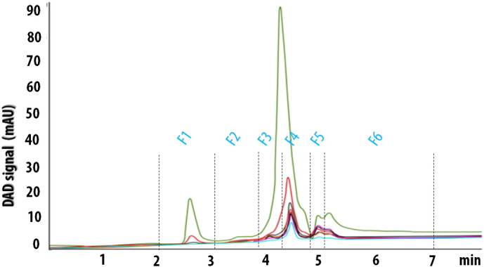
HPLC chromatogram shows retention times (minutes) of the six separated fractions of the A. fulica mucus. The signal according to the diode array detector (DAD) is represented in milli-arbitary unit (mAU). The fractions were scanned at different ultraviolet wavelengths; 210 (pink), 220 (green), 230 (purple), 250 (red), 280 (brown green) and 290 (blue) nm.