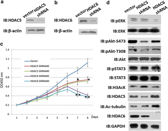
Fig. 2.

Knockdown of HDAC5 or HDAC6 inhibited the proliferation of A375 cells. The stable cell line of A375 cells with HDAC5 or HDAC6 knockdown were constructed using shRNA primers. a and b Western blotting was used to detect HDAC5 or HDAC6 expression in A375 cells. β-actin was used as an internal control. c CCK8 was used to count the cell number of stably knocked down HDAC5 or HDAC6 in A375 cells. Cell viability was measured using the Cell Counting Kit-8 (CCK-8, Dojindo Laboratories, Kumamoto, Japan) according to the manufacturer’s instructions. Transiently transfected cells were seeded in a 96-well plate and then cultured at 24-hour intervals for 5–7 days. Cell viability was then measured using the CCK-8 assay. Absorbance was measured at 450 nm as an indicator of cell viability. All experiments were independently repeated at least three times. *p value <0.01, **p value <0.001. p value <0.05 was considered as significant differences. d Western blotting was used to detect the signaling pathway related to proliferation. Acetylated-Histone H3 and acetylated-a-tubulin were used as control for monitoring HDAC5 and HDAC6 knocking down results, respectively. The antibodies used for western blotting are listed in Methods
