
Figure 2.
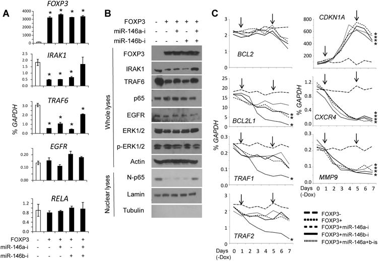
FOXP3-miR-146-NF-κB axis in breast cancer cells. A. Quantification of FOXP3, IRAK1, TRAF6, EGFR, and RELA mRNA levels at 72 hours after FOXP3 induction. Data are presented as the mean and SD of triplicates. All *, p < 0.05, two-tailed t test. B. Representative western blots detecting the expression of FOXP3, IRAK1, TRAF6, EGFR, ERK1/2, p-ERK1/2, p65, and nuclear p65 (N-p65) 72 hours after FOXP3 induction. C. Quantification of NF-κB target gene mRNA expression after FOXP3 induction. Down arrows indicate transfection with scramble miR or miR-146a/b inhibitors. *, p < 0.05, Fisher's PLSD test. All experiments were repeated three times.