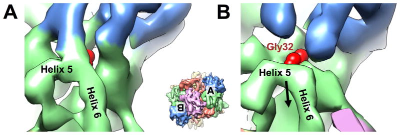
Figure 4. Binding of PhnK causes Gly32 of PhnJ more exposed.
(A) On the side without PhnK bound (apo side), the Helix 5 of PhnJ connects PhnH. (B) On the side with PhnK bound (bound side), the densities for H5 and H6 of PhnJ moves away from PhnH to expose the Gly32 of PhnJ. The density map of PhnG2H2I2J2K is rendered at 4.5 s above mean and colored coded as in Figure 2. The red sphere model denotes Gly32 of PhnJ. Inset shows the map of PhnG2H2I2J2K in the same orientation as Figure 2E with the locations of (A) and (B) labeled. See also Figure S4.

