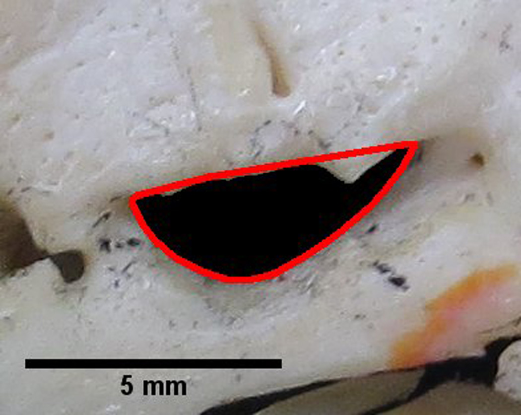
Figure 1.

Foramen ovale outlined by a red convex hull. The solidity of the foramen can be determined by dividing the area within the foramen by the area contained within the convex hull. A perfectly convex shape has a solidity of 1. This image illustrates a foramen with a solidity of 0.87. Therefore, certain parts of the foramen outline are convex. These convexities may indicate bony structures projecting into the foramen, which can be visualized in the figure. The measurement of solidity may aid in identification of bony projections encroaching upon the foramen.
