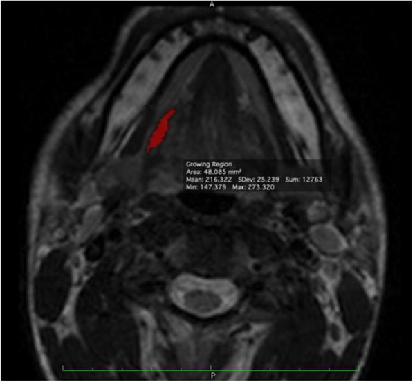
Figure 3.

Hyoglossus segmentation example. At one slice level, the completed segmentation of the hyoglossus on the right side of the subject will appear as shown. The voxel selections made by the semiautomated muscle segmentation algorithm in Osirix were accepted or rejected until the final segmentation fit distinct muscle boundaries in the axial T2-weighted MR image series.
