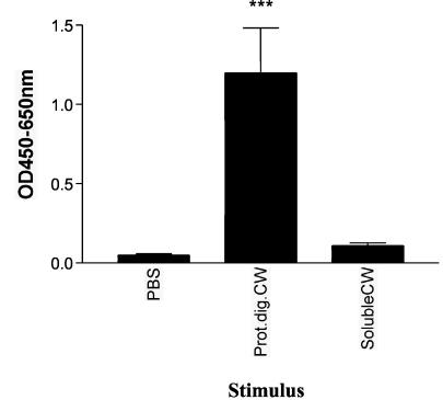
FIG. 4.
Bovine γδ T cells produce IFN-γ following culture with proteolytically digested mycobacterial cell wall (Prot.dig.CW). Monocytes and γδ T cells were isolated from the peripheral blood of 10 animals by using magnetic beads. Monocytes and γδ T cells from each individual animal were incubated with proteinase K-treated M. bovis cell wall, the SDS-soluble fraction of M. bovis cell wall, or PBS for 48 h. Supernatants were then tested for the presence of IFN-γ. Data are presented as mean OD at 450 to 650 nm ± standard error from two successive experiments with five animals per experiment. Significant IFN-γ production was determined by one-way ANOVA with the Tukey posttest. ***, P < 0.001.

