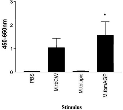
FIG. 5.
IFN-γ production by γδ T cells following stimulation with mycobacterial mAGP but not with mycobacterial lipids. Monocytes and γδ T cells were isolated from the peripheral blood of five animals by using magnetic beads. Monocytes and γδ T cells were incubated with M. tuberculosis cell wall (M.tbCW), lipid extract from M. tuberculosis, M. tuberculosis mAGP, or PBS for 48 h. Supernatants were frozen and then tested for the presence of IFN-γ. Data are presented as mean OD at 450 to 650 nm ± standard error for five animals. Significant IFN-γ production was determined by one-way ANOVA with the Tukey posttest. *, P < 0.05.

