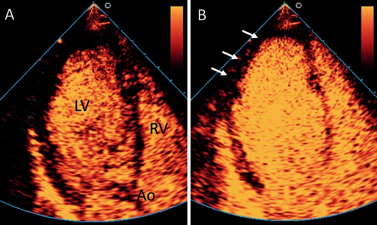
Fig. 2.
Assessment of myocardial perfusion using CEUS. Example of an abnormal myocardial perfusion echocardiogram. a Apical three chamber view. After administration of the contrast agent, a high mechanical index flash is given to destroy the contrast agent that is present in the myocardium. Thereafter, the left ventricular myocardium does contain no or only a limited amount of contrast agent. Ao aorta, LV left ventricle, RV right ventricle. b After a short period, the myocardium is filled with blood and contrast agent. There is an apical and lateral perfusion defect visible (arrows), indicating a significant coronary stenosis. Example reproduced from [84]

