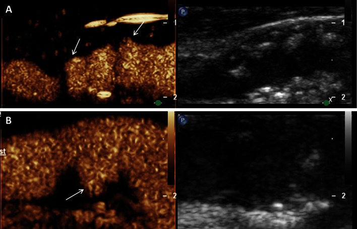
Fig. 3.
Assessment of vessel wall irregularities and plaque ulcerations on carotid artery using CEUS. a Mixed hypo- and hyperechoic plaques at the carotid bulb on B-mode ultrasound (right side) and CEUS imaging (left side) with surface irregularities (arrows). b Plaque ulceration (arrow) on CEUS imaging (left side) at the origin of the internal carotid artery not detected on B-mode ultrasound (right side)

