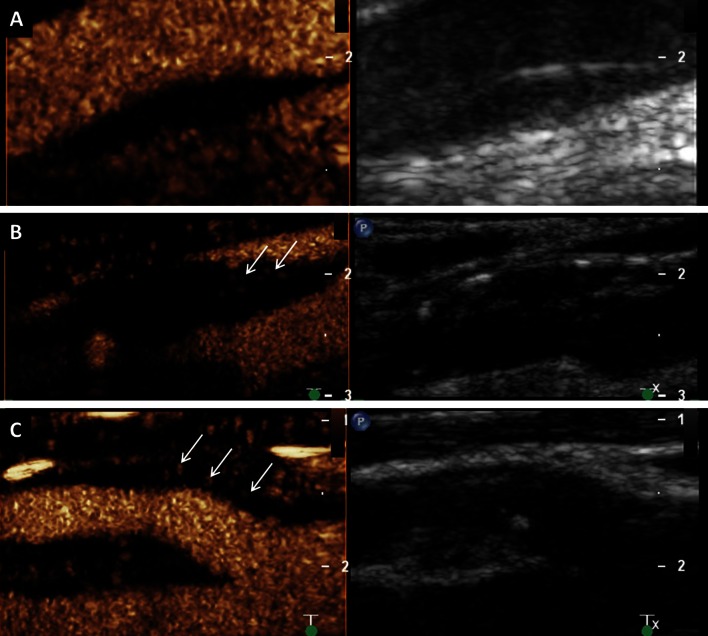
Fig. 4.
Visual based grading of intraplaque neovascularization on CEUS imaging. a No enhancement: Small plaque on the fare wall of the internal carotid artery on B-mode ultrasound (right side) without intraplaque neovascularization on CEUS imaging (left side). b Moderate enhancement: Mixed hypo- and hyperechoic plaques at the carotid bulb on B-mode ultrasound (right side) and CEUS imaging (left side) with moderate intraplaque neovascularization on the plaque shoulder (arrows). c Extensive enhancement: Hypoechoic plaque at the origin of the internal carotid artery on B-mode ultrasound (right side) and CEUS imaging (left side) with extensive intraplaque neovascularization including the plaque core (arrows)

