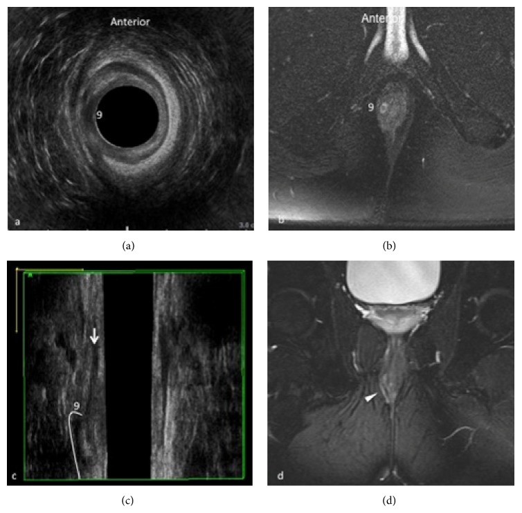
Figure 3.
Intersphincteric fistula at 9 o'clock. 3D-EAUS demonstrates the proximal origin of the fistulous tract from the internal anal sphincter and its location in the intersphincteric plane on both axial (a) and coronal plane (c), better depicting the fistulous tract in the intersphincteric space than MRI (b, d).

