
Figure 4.
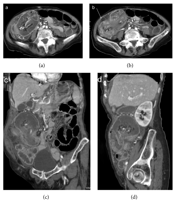
Enterocolic (ileocaecocolonic) intussusception due to a caecal carcinoma. CT images on axial scans (a, b) and oblique reformatting (c, d) show lymph nodes and vascular engorgement in the intussuscepted mesentery and fluid distention of the intussuscipiens. Extraparietal air indicates local perforation (arrow).