Figure 5.
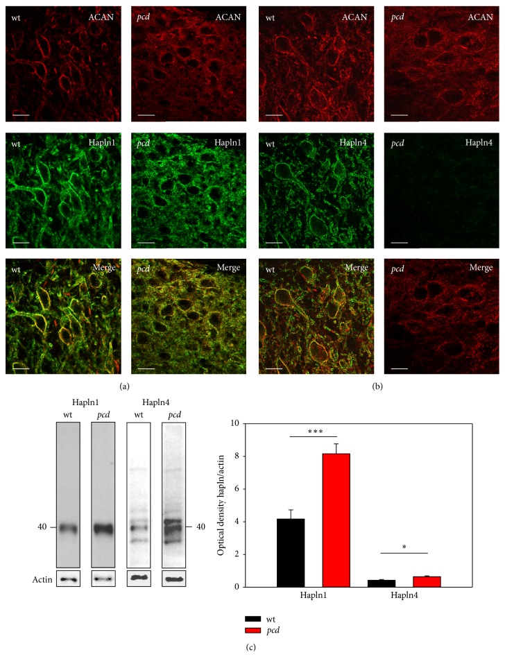
Comparison of link protein expression in DCN. DCN neurons are visualized by aggrecan immunoreaction (red). (a) Hapln1 labeling (green) surrounds the DCN neurons in both genotypes, matching the aggrecan immunoreactivity; additionally in pcd hapln1 immunoreaction is distributed throughout the whole parenchyma. (b) Hapln4 (green) encloses the DCN neurons in wt mice. In contrast, hapln4 in pcd exhibits virtually no immunoreaction. Scale bar: 20 μm. (c) Western blot reveals protein bands at approximately 40 kDa for link proteins. Quantification of the link proteins yielded an elevated protein level of both components in pcd (hapln1 p < 0.01; hapln4 p < 0.05). Data are given as mean ± SEM.

