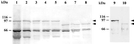
FIG. 2.
Immunoblot analysis of Ami molecules produced by serovar 1/2 and 4b strains. Blots prepared from SDS bacterial extracts were probed with anti-InlB (lanes 1 to 8) or anti-Ami (lanes 9 and 10) polyclonal antibodies. Lane 1, EGD as a control serovar 1/2a strain; lane 2, LO28 (serovar 1/2c); lane 3, ATCC 19111 (serovar 1/2a); lane 4, CNL 880203 (serovar 1/2b); lane 5, CHUT 861141 (serovar 1/2c); lane 6, CHUT 82337 (serovar 4b); lane 7, CHUT 850212 (serovar 4b); lane 8, INRA 76 (serovar 4b); lane 9, EGD (serovar 1/2a); lane 10, CHUT 82337 (serovar 4b). Note that the main band, corresponding to the complete form of Ami (arrowheads), is at ∼85 kDa in serovar 4b strains and at ∼100 kDa in serovar 1/2 strains. The ∼65-kDa band present in all extracts probed with anti-InlB antibodies is InlB.

