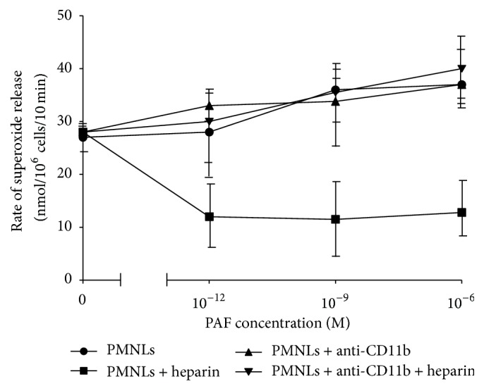
Figure 7.

The effect of anti-CD11b on superoxide release in the presence of heparin. Superoxide release from NC PMNLs preincubated with increasing concentrations of PAF and IC-PE antibodies (●). In addition, superoxide release from NC PMNLs was also measured after 15 min preincubation with PAF and anti-CD11b-PE antibodies (▲). Furthermore, cells were washed and subjected to 30 min incubation with 25 U/mL of heparin (■). The rate of superoxide release from NC PMNLs was also measured after preincubation of NC PMNLs with PAF + anti-CD11b antibodies, before heparin incubation (▼). The rate of superoxide release was determined with 0.32 × 10⁻⁷ M PMA-stimulated PMNLs. The changes in optical density were monitored at 549 nm continuously in the presence of 0.08 mM cytochrome C. Data are represented as nmoles/106 cells/10 min; n = 3.
