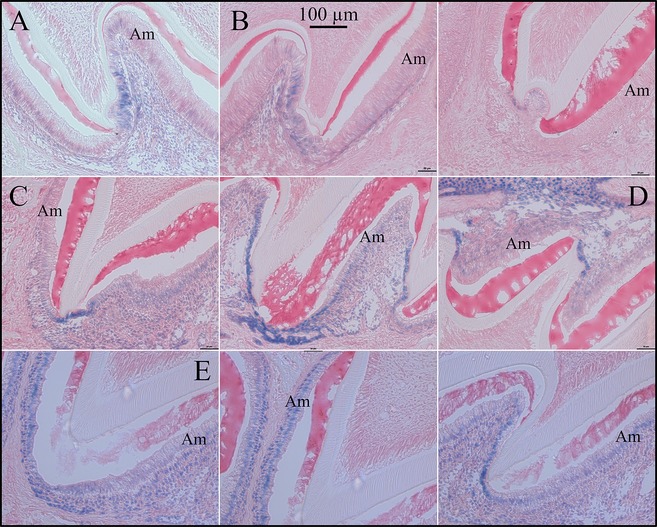
Figure 8.

X‐gal Histostaining of Fam83h‐knockout/NLS‐lacZ‐knockin (null) developing molars. A: Cusp region of Fam83h null D5 maxillary second molar showing positive staining in epithelia covering the enamel‐free area. The first molar and incisor ameloblasts were negative or trace for X‐gal staining (Figs. S13–S14). B: Cusp region of Fam83h null D6 maxillary second molar showing weak/positive staining in epithelia covering the enamel‐free area. No other ameloblasts were positive in D6 molars or incisors (Fig. S14). C: Cusp region of Fam83h null D9 maxillary second molar showing positive staining in epithelia covering the enamel‐free area and weak staining in maturation stage ameloblasts (Fig. S15). D: Cusp region of Fam83h null D9 mandibular second molar showing positive staining in epithelia covering the enamel‐free area. E: Day 11 maxillary molars show positive histostaining for maturation stage ameloblasts.
