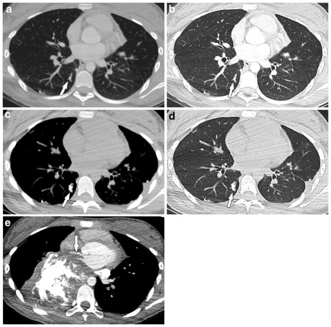
Fig. 7.
A 12-year-old girl with osteosarcoma of the right proximal humerus with metastasis to the lungs and tumor thrombus in the pulmonary artery, pulmonary vein and left atrium. a, b Initial and (c, d) 4-month follow-up axial post-contrast chest CT scans in mediastinal and lung windows show a progressive increase in mineralization and beaded appearance of the right lower lobe distal pulmonary artery (arrows). Surgery was refused and the tumor progressed on chemotherapy. e Chest CT scan with contrast agent in mediastinal windows 22 months later shows significant progression of metastatic disease with extension into the right inferior pulmonary vein and left atrium (arrow)

