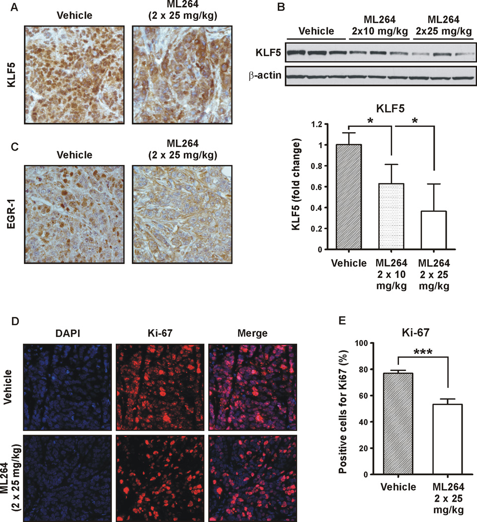
Figure 7. ML264 treatment reduced the expression levels of KLF5 and EGR1 in DLD-1-derived tumor xenografts.
(A) Representative immunohistochemistry staining of KLF5 in DLD-1-derived xenografts treated with vehicle (left panel) and treated with ML264 twice per day at 25mg/kg (right panel) for ten days. (B) Western blot analysis (top panel) and the quantitative analysis (bottom panel) of KLF5 levels in DLD-1-derived xenografts treated with vehicle and treated with ML264 twice per day at 10mg/kg and 25mg/kg for ten days. Results shown from three independent experiments. Data represent mean ± S.D. (n=3). *p < 0.05. (C) Representative immunohistochemistry staining of EGR1 in DLD-1-derived xenografts treated with vehicle (left panel) and treated with ML264 twice per day at 25mg/kg (right panel) for ten days. (D) Immunofluorescence staining of Ki-67 (proliferative marker). Top panel – vehicle treated mice, Bottom panel – ML264-treated mice. (E) Quantitative representation of (D). Three fields were counted for each condition. Data represent mean ± S.D. (n=3). ***p < 0.001.

