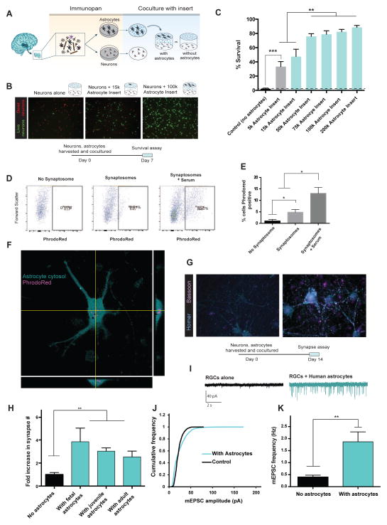
Figure 2.
Functional characterization of human astrocytes. A. Schematics of co-culture experiments. 17–20 gestation week fetal human astrocytes and neurons were purified by immunopanning, grown in the same wells separated by porous inserts. B. Calcein stain of live neurons (green) and ethidium homodimer stain of dead neurons (red) in the presence and absence of astrocytes. Scale bars: 100μm C. Quantification of survival rate. N=7–8 images per condition. Data represent mean ± SEM in all the figures unless otherwise noted. **, p<0.01. ***, p<0.001. Two-tail unpaired T-test was used for data in C, H, and K. D. Human astrocytes engulf synaptosomes in vitro. FACS plot of human fetal astrocytes incubated without synaptosomes, with synaptosomes, or with synaptosomes and 5% serum. E. Percentages of synaptosome-positive astrocytes. *, p=0.05, one-tailed Wilcoxon rank sum test. n=3 patients. F. Confocal image of a human fetal astrocyte stained with Vibrant CFDA (cyan) engulfing PhrodoRed labeled synaptosomes (magenta) Scale bar: 20μm. G. Retinal ganglion cells form more synapses in the presence of human astrocytes. Cyan: immunofluorescence of post-synaptic marker, Homer. Magenta: immunofluorescence of pre-synaptic marker, Bassoon. Scale bar: 10μm. H. The number of synapses (Homer/Bassoon double positive puncta) in the presence and absence of human astrocytes. N=10–20 images per patient. Each image contains on average two cells. Fetal: 1 patient, age: 18 gw. Juvenile: 3 patients, age: 8, 13, 18 yo. Adult: 3 patients, age: 26, 41, 47 yo. **, p<0.01. I. Representative traces of whole-cell patch clamp recordings from RGCs cultured with or without feeder layers of human astrocytes in the presence of TTX. Few mEPSCs were observed without feeder layers of human astrocytes. J. Human astrocytes significantly increased the amplitude of mEPSCs (p=.0001 Kolmogorov-Smirnov test, n= 488 and 2837 mEPSCs from 16 and 18 cells for no astrocyte and with astrocytes respectively). K. The frequency of mEPSCs was significantly increased in the presence of human astrocytes (Two-tailed Wilcoxon rank sum test. p=0.004. n = 16–18 neurons per condition).

