Figure 2.

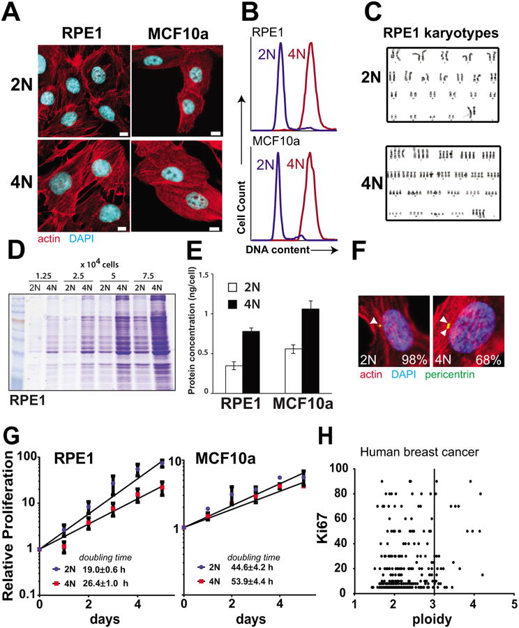
Matched pairs of diploid-polyploid cells were generated from epithelial breast cell types. A. Representative images of diploid (2N) and polyploid (4N) cells derived from RPE1 and MCF10a cell lines. Scale bar = 10 μm. B. Flow cytometry demonstrating double DNA content of tetraploid cells after staining with propidium iodide. C. Karyotype of diploid and polyploid matched RPE1 cells. D-E. Protein levels are higher in tetraploid cells relative to diploid as determined by coomassie staining of extracts (D) and Bradford Assay (E). F. Polyploid RPE1 cells have supernumerary centrosomes. Cells were synchronized using aphidicolin and stained. Pericentrin foci are marked with arrows and percentage of cells that harbor the indicated number of centrosomes is indicated (n>40). G. 4N cells proliferate more slowly than 2N cells. Cell number was quantified in proliferating 2N and 4N cells and doubling time was calculated. H. High ploidy in human breast cancer samples does not correlate strongly with the Ki67 proliferation marker.
