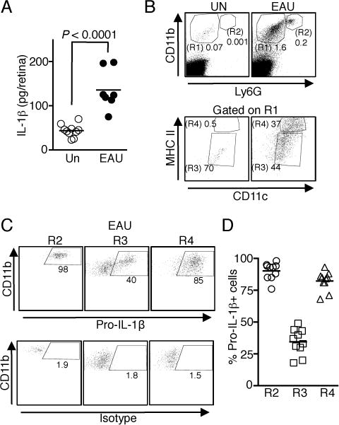
FIGURE 1.

IL-1β is expressed in the retina during EAU. Retinae of WT mice without immunization (Un) and with EAU were isolated on day 21 post-immunization, digested with collagenase and analyzed by ELISA (A) or flow cytometry (B–D). (A) Analysis of IL-1β secretion by ELISA. Data are from 2 independent experiments with a total of 7–10 mice. (B) Macrophages/DCs (CD11b+ Ly6G−, R1) and neutrophils (CD11b+ Ly6G+, R2) that had infiltrated into the retina were first identified by CD11b and Ly6G staining. Macrophages and DCs were defined as CD11c+ MHC IIlow macrophages (R3) and CD11chi MHC IIhi DCs (R4) (C and D) Expression of intracellular pro-IL-1β in the immune cells identified in Fig. 1B after EAU induction. Shown are representative flow cytometric plots (C) and as well as data from 10 individual mice (D).
