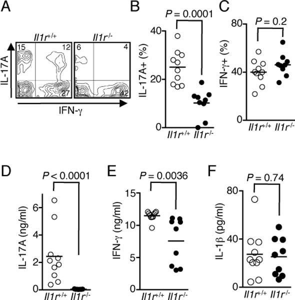
FIGURE 4.

IL-1R-dependent signaling is required for Th17 cell differentiation. (A–C) EAU was induced in WT and Il1r−/− mice, retinae were isolated on day 21 post-immunization and analyzed by the intracellular cytokine staining assay. Shown are representative flow cytometric plots (A) and percentages of CD4+ T cells expressing IL-17 (B) and IFN-γ (C) from individual mice. (D and E) Cervical and draining lymph node cells were re-stimulated with IRBP1–20 peptide for 3 d. Amounts of IL-17 (D), IFN-γ (E), and IL-1β (F) in the culture were determined by ELISA. Data shown are from 2 independent experiments with a total of 10 mice/group.
