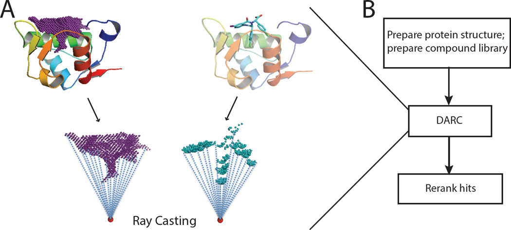
Figure 1. Docking Approach using Ray-Casting.
(A) DARC first casts a set of rays emanating from an origin within the protein (red dot), and maps the topography of the surface pocket by monitoring intersection of these rays with the pocket (left). To evaluate the shape complementary of a given ligand for this pocket, DARC casts the same rays (from the same origin) and monitors their intersection with the ligand (right). If ligand (in its current position and orientation) is perfectly shape-complementary to the pocket, each ray will intersect the ligand at the same distance from the origin as it intersected the protein surface pocket. By moving the ligand to maximize this shape complementarity, a ligand can be docked into a protein surface pocket. (B) A schematic diagram of the complete workflow split into three stages: pre-DARC preparation, DARC, and post-DARC re-ranking/filtering of the docked models.

