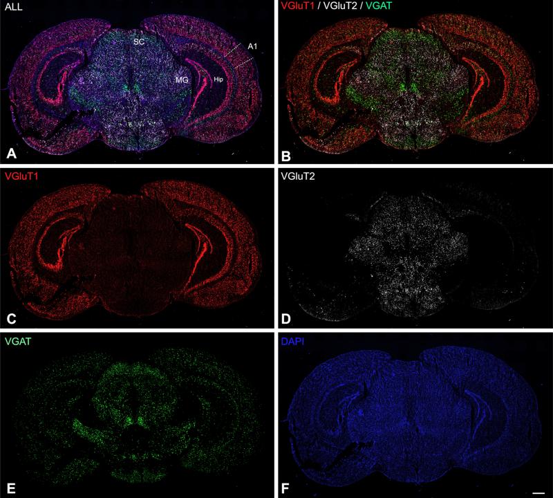
Fig. 6.
Triple FISH assay in coronal sections of adult mouse at the level of A1 and MGB. The low-magnification image montages obtained at 10× show combined (a) and single channel expression (b–f) for VGAT, VGluT1, and VGluT2 mRNA, counterstained by DAPI. SC superior colliculus, Hip hippocampus, MG medial geniculate body. Scale bars 500 μm in all panels

