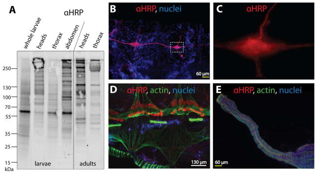
Figure 1. In vivo detection of core α1,3-fucosylated N-glycan epitopes using anti-HRP antibody.
(A) Dissected tissues of Anopheles gambiae larvae and female adults as well as whole larvae were subject to Western Blot analysis using anti-HRP antibody (αHRP) which demonstrates a particular concentration of the α1,3-fucose epitope in the adult head; Coomassie staining (not shown) indicated comparable amounts of protein for the samples shown. (B–E) Female adult abdomens were further dissected into dorsal and ventral halves prior to immunofluorescence staining with anti-HRP. (B) Anti-HRP staining of the ventral nerve cord and ganglia from two abdominal segments; nuclei of surrounding fat body, muscle cells and haemocytes can be seen in the blue channel (Hoechst 33342). (C) High magnification image of inset from panel B, showing the increased magnitude and seemingly punctate anti-HRP reactivity in the concentrated nervous tissue of the ganglion. (D) Anti-HRP staining of pericardial cells (nephrocytes) along with muscular staining of the attached mosquito heart (at top) and some of the ventral abdominal muscles (at bottom); actin stained with phalloidin (in green). (E) Anti-HRP reactivity with nervous tissue attached to the mosquito midgut (muscles shown in green); in this instance anti-HRP reactivity was observed on a finer scale than seen in panels A–C.

