. 2015 Dec 22;31(4):188–197. doi: 10.5625/lar.2015.31.4.188

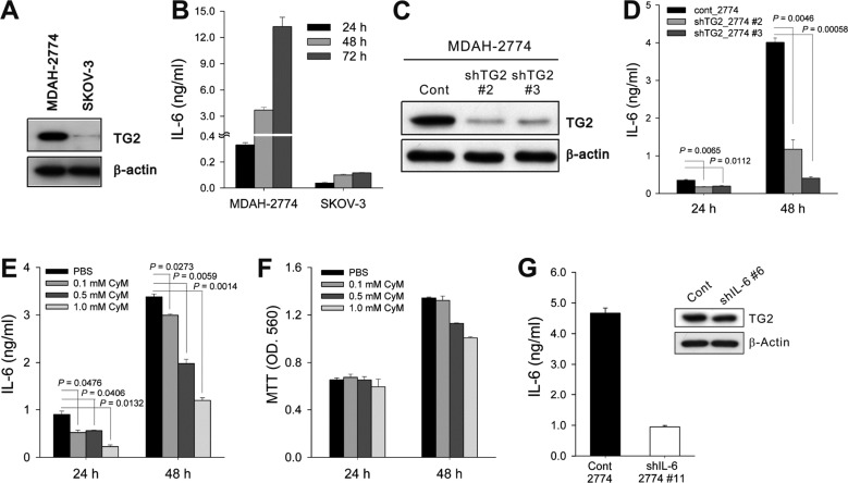
Figure 1. TG2 expression levels in cancer cells correlated with IL-6 production. (A) TG2 expression in the two human ovarian cancer cell lines was analyzed by Western blotting. (B) IL-6 levels in culture supernatants of ovarian cancer cells were determined by enzyme-linked immunosorbent assay (ELISA). Cells (1×10⁴) were cultured in a 24-well tissue culture plate, and the supernatants were collected 24, 48, and 72 h after culture. (C) MDAH-2774 human ovarian cancer cells (cont_2774, control empty-vector transfected; shTG2_2774, TG2-knocked-down) were stably transfected with short hairpin RNAs (shRNAs) targeting TG2. The efficiency of knockdown was assessed by Western blotting. (D) IL-6 levels in culture supernatants of control and TG2-knocked-down MDAH-2774 cells were determined by ELISA. Cells (1×10⁴) were cultured in a 24-well tissue culture plate, and the supernatants were collected 24 and 48 h after culture. (E, F) The effect of the TG2 inhibitor cyteamine (CyM) on IL-6 secretion (E) and cell viability (F) in control MDAH-2774 cells was determined by ELISA and the MTT assay, respectively. (G) MDAH-2774 cells were stably transfected with shRNAs targeting IL-6. The efficiency of knockdown was verified by measuring IL-6 levels in the culture supernatants. TG2 expression of control and IL-6-knocked-down MDAH-2774 cells was analyzed by Western blotting. Data represent the mean±standard deviation (SD), based on three independent experiments using samples from triplicate cell cultures.

Figure 1