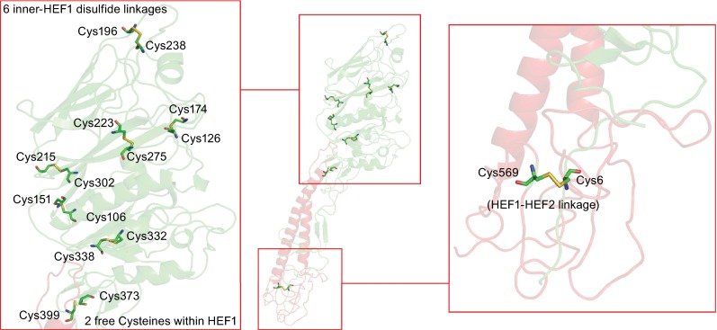
Figure 5.

Location of intramolecular disulfide linkages in HEF. The middle part shows the secondary structure of a HEF monomer. HEF1 and HEF2 subunits are drawn in red and green, respectively. The left part shows the head domain which contains six disulfide linkages and also two free cysteines. The right part shows the location of the only disulfide linkage between HEF1 and HEF2. The figure was created with PyMol from PDB file 1FLC. HEF1 and HEF2 subunits are drawn in red and green, respectively
