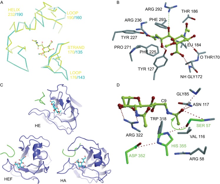
Figure 8.

Structures of receptor binding site and esterase site of HEF, HA and HE. (A) Superimposing of HEF and HA receptor binding sites complexed with ligand (9-acetamidosialicacid α-methyl glycoside). The yellow-green lines and the light blue lines represent the binding elements of HA and HEF, respectively. (B) Structure of HEF binding sites complexed with the receptor. Key residues forming hydrogen bonds with the ligand are shown. (C) Comparison of the receptor binding topology of the hemagglutinin-esterase (HE) protein of coronavirus, HEF and HA with their ligands. The bound ligands are αNeu4,5,9Ac32Me in HE, αNeu5,9Ac22Me in HEF and αNeu5Ac2Me in HA are shown in stick representation. The ligand is bound to HE in an opposite orientation compared to HEF and HA. (D) Structure of the esterase active site of HEF. Key residues forming hydrogen bonds with the ligand are shown. (A), (B) and (D) were taken from reference (Rosenthal et al., 1998) and (C) from reference (Zeng et al., 2008) with permission
