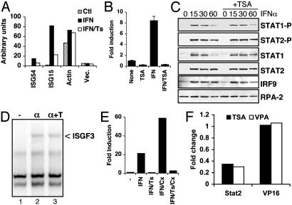
Fig. 2.
HDAC function is required for transcription without affecting Janus kinase–Stat signaling. (A) HeLa cells were untreated or treated with IFNα with or without TSA for 1 h, before isolation of nuclei for in vitro run-on transcription reactions. Labeled RNA was hybridized with filter-bound cDNA for the indicated sequences and quantified by PhosphorImager analysis. (B) HeLa cells transfected with ISG54-luciferase together with CMV-LacZ were treated with TSA and IFNα, as indicated, and cell extracts were analyzed for luciferase activity, normalized to β-galactosidase activity, and reported as fold induction over untreated cells. (C) HeLa cells were untreated or treated with IFNα in the absence or presence of TSA, as indicated, before isolation of nuclear extracts. Western blots were probed with antibodies against phospho-Stat1 and Stat2, total Stat1 and Stat2, IRF9, and RPA-2, as indicated. (D) Extracts from HeLa cells treated with IFNα with or without TSA were analyzed for the presence of ISGF3 by electrophoretic mobility-shift assay. (E) ISG54 transcript levels were quantified from HeLa cells treated with IFNα, TSA, and Cx, as indicated. (F) HeLa cells were transfected with UAS-luciferase along with Gal4 DNA-binding domain alone or fused with the transactivation domain of Stat2 or VP16. Fold change in luciferase activity in the presence of TSA or VPA relative to untreated samples is shown.

