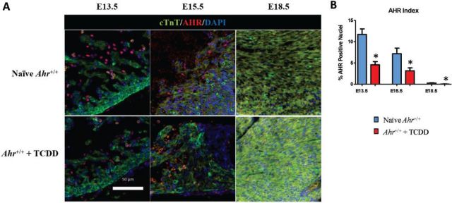
FIG. 1.
AHR localization during early heart formation. A, Five-micrometer sections of embryonic hearts collected at E13.5, E15.5, and E18.5 from Ahr+/+, naïve or exposed to AHR ligand (TCDD) in utero, were used for immunofluorescent detection of AHR (red) and cardiac troponin T (green). Staining with DAPI identifies the nuclei. AHR, aryl hydrocarbon receptor; cTnT, cardiac troponin T; DAPI, nuclei; scale bar = 50 µm. B, Quantification of nuclear localization of AHR (AHR Index) at the indicated embryonic developmental times. The AHR index is shown as the mean percent positive nuclei ± SEM; *P ≤ .05. Full color version available online.

