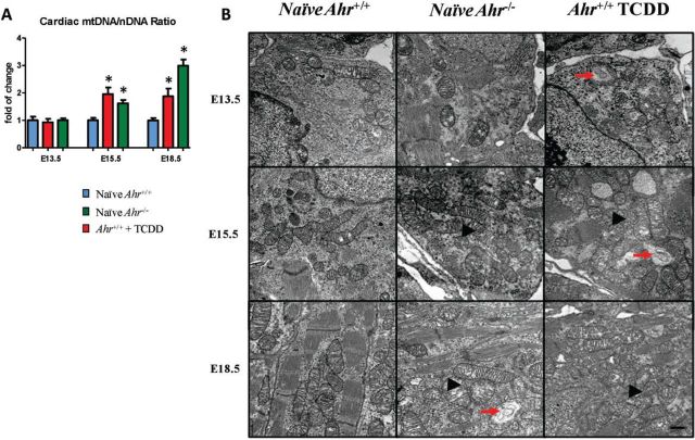
FIG. 7.
Disruption of AHR signaling induces changes in mitochondria abundance and structure. A, Quantification of heart mitochondrial DNA relative to nuclear DNA, defined as the ratio of mtDNA to nDNA ratio at E13.5, E15.5, and E18.5 and expressed as the mean fold change relative to naïve Ahr+/+ hearts ± SEM; *P ≤ .05. B, Ultra-thin sections of embryonic hearts collected at E13.5, E15.5, and E18.5 from Ahr−/− and Ahr+/+, either naïve or exposed to ligand in utero, were used for transmission electron microscopy evaluation of the embryonic heart ultrastructure. Representative photomicrographs illustrating higher density of mitochondria (arrowheads) within the embryonic cardiomyocyte sarcoplasm in Ahr−/− and in ligand-exposed Ahr+/+ compared with naïve Ahr+/+. Individual or clusters of mitochondria showed ultrastructural features of stress and degeneration (arrows), as evidenced by focal to global swelling, loss of matrix density, as well as cristae unpacking, disorganization, and cristolysis, affecting a higher number of mitochondria in AHR ligand-exposed Ahr+/+ hearts. Uranyl acetate and lead citrate stain. Scale bar = 500 nm.

