Figure 6.
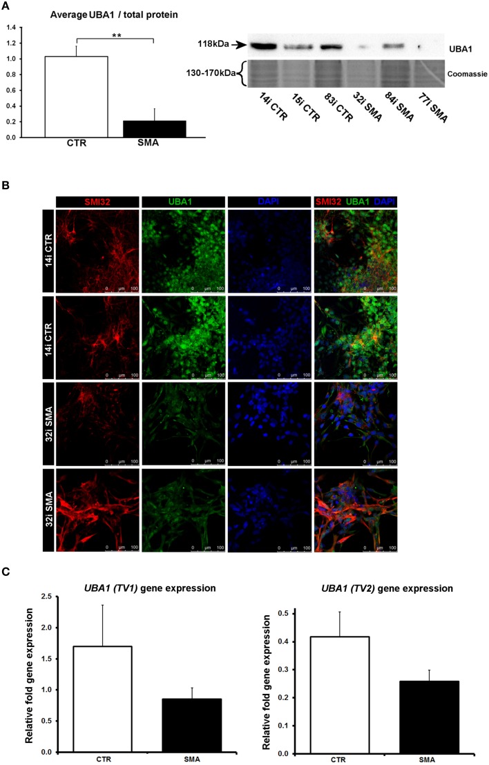
Reduction and differential localization of UBA1 in SMA motor neurons. (A) Representative western blot showing UBA1 protein levels in three different control and SMA motor neuron cell lines, along with Coomassie stained gel as loading control. The graph illustrates the average integrated density of the UBA1 bands from this blot/total protein (Coomassie gel), as determined by ImageJ software. Error bars represent standard error from the mean and statistical significance was calculated using an unpaired, one-tailed t-test with two-sample unequal variance. (B) Representative confocal images indicate a reduction of UBA1 levels in SMI32 positive 32i SMA motor neurons and mostly cytoplasmic distribution (in comparison to the mostly nuclear distribution seen in the 14i control (CTR) motor neurons). (C) Average gene expression levels of UBA1 transcript variants 1 (TV1) (p = 0.14; unpaired, one-tailed t-test) and 2 (TV2) (p = 0.08; unpaired, one-tailed t-test) in the control and SMA motor neuron cell lines [the same six patient and control cell lines shown in (A)], as determined by qRT-PCR. Relative fold expression was normalized to H9 hESCs. Error bars represent standard error from the mean. CTR, control; MNs, motor neurons. **p ≤ 0.01.

