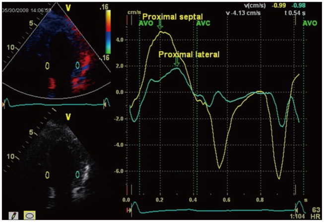
Fig. 2. Derived tissue Doppler recordings from the proximal septum and lateral wall are shown for a patient with moderate to severe chronic AR + PEF. The time to reach peak velocity was 110 msec later for the proximal lateral wall demonstrating systolic dyssynchrony. AR + PEF: chronic aortic regurgitation and preserved left ventricular ejection fraction.

