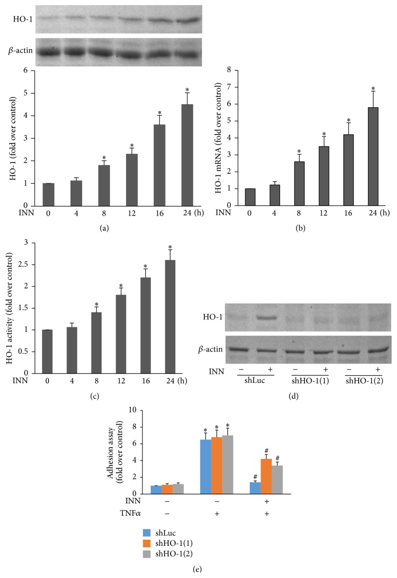
Figure 2.
The role of HO-1 in INN suppression of TNFα-triggered HL-60 cell adhesion. ECs were incubated with 10 μM INN for the indicated time points. Aliquots of lysate (50 μg) were subjected to Western analysis for HO-1 protein expression (a). Total RNA was extracted from ECs and underwent real-time PCR with specific primers for HO-1 and β-actin (b). HO-1 activity was measured three times in each treatment group as mentioned in Methods. Data are expressed as fold increase of HO-1 activity versus control (c). ECs transfected with shHO-1 were incubated with 10 μM INN for 24 h prior to being stimulated with 1 ng/mL TNFα for additional 6 h (d). (e) ECs were incubated with 1 ng/mL TNFα in the presence or absence of INN for 8 h. Adhesion assay was performed as described in Materials and Methods. Values are means ± SD (n = 4). ∗ P < 0.05 versus control; # P < 0.05 versus TNFα.

