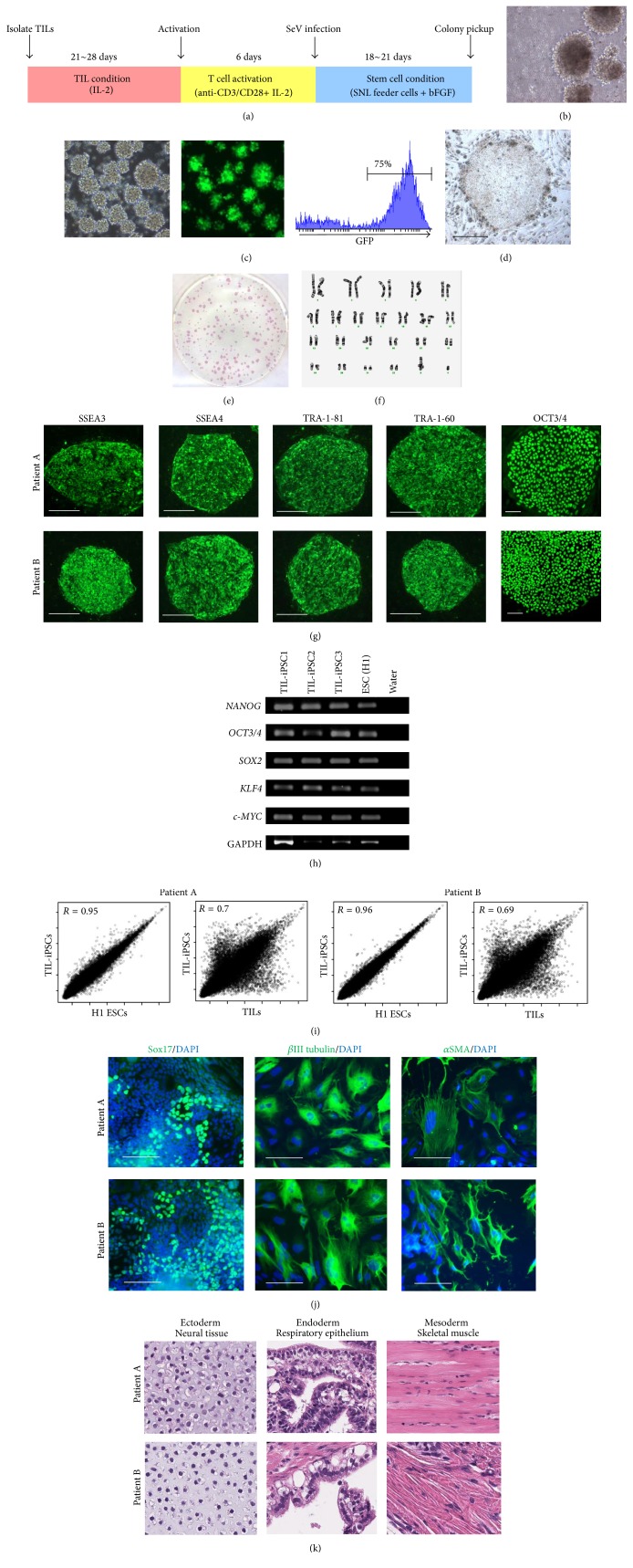
Figure 2.
Generation of iPSCs from melanoma tumor-infiltrating lymphocytes. (a) Time schedule outlining expansion, activation, and reprogramming of TILs to generate iPSCs. (b) Morphologies of TILs when they started to expand 2-3 weeks after initiation of culture. (c) Efficient GFP introduction by Sendai virus (SeV) in TILs transfected at a MOI of 20. (d) Typical ESC-like iPSC colonies on day 21 after SeV infection. (e) Examples of 6-well plate containing SeV-reprogrammed iPSC clones stained for alkaline phosphatase (ALP), showing numerous ALP-positive colonies. (f) Cytogenetic analysis was performed on twenty G-banded metaphase cells from one of TIL-derived iPSCs (TIL-iPSCs). All twenty cells demonstrated a normal karyotype. (g) Immunofluorescence staining for pluripotency and surface markers (SSEA3, SSEA4, TRA-1-81, TRA-1-60, and OCT3/4) in iPSCs derived from melanoma TILs. Scale bars represent 100 μm. (h) RT-PCR analysis for the human ES cell marker genes NANOG, OCT3/4, SOX2, KLF4, and c-MYC in TIL-iPSCs and ESCs (H1). (i) Scatter plots comparing the global gene expression profiles of TIL-iPSCs and ESCs and TIL-iPSCs and TILs. (j) Immunofluorescence staining for SOX17 (endodermal marker), βIII tubulin (ectodermal marker), and αSMA (mesodermal marker) in TIL-iPSC-derived differentiated cell. Nuclei were counterstained with DAPI. Scale bars represent 100 μm. (k) Hematoxylin and eosin-stained representative teratoma sections of TIL-iPSC clones from patients (A) and (B) (6 weeks after injection into NOD/SCID mice).

