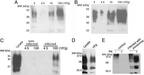
Fig. 1.
The cell culture medium of infected cells contains infectious PrP. (A) The culture medium from 5 × 10⁶ infected Rov cells was submitted to differential centrifugation (11). The medium was submitted to a first centrifugation for 5 min at 4,500 × g to remove cells in suspension. The supernatant was ultracentrifuged at 10,000 × g for 30 min to remove cell debris, and, finally, the last supernatant was ultracentrifuged at 100,000 × g for 1 h. The resulting pellets of each centrifugation step were analyzed by Western blot for PrP. (B) The culture medium of 5 × 10⁶ infected Mov cells was analyzed for PrP as in A. (C) After 5 days of culture, the media of 2 × 10⁷ control Mov cells or scrapie-infected Mov cells were harvested and centrifuged for 5 min at 4,500 × g to remove potentially dissociated cells; the supernatant was then recentrifuged at 100,000 × g for 1 h. The pellets were digested with PK and analyzed for the presence of PK-resistant PrPsc by Western blotting with SAF 84. The lysate comprised Mov cells. (D) The medium of 3 × 10⁷ infected Rov cells was centrifuged and analyzed as in C. The lysate comprised Rov cells. (E) Cell culture medium from 3 × 10⁷ infected Rov cells was ultracentrifuged as above. The pellet was resuspended and incubated for 7 days with uninfected Rov cells. Cultures were grown for several weeks and monitored for the accumulation of cell-associated PK-resistant PrP. Total PrP was obtained by methanol precipitation of undigested cell lysates (lanes 1 and 3, 25 μg of proteins). PrP was isolated from PK-digested cell lysates (lanes 2 and 4, 250 μg of proteins). MW indicates molecular mass in all figures.

