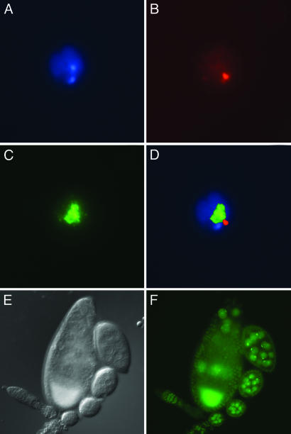
Fig. 5.
POF localization in S2 cells and in ovaries. (A-D) S2 cells: DAPI (A), anti-POF (B), anti-MSL3 (C), and merged (D). The merged image shows that POF staining is separable from the MSL3-labeled X chromosome. Immunofluorescence of POF.EYFP as detected in live ovary preparations. (E) Nomarski image. (F) EYFP fluorescent image. The strong signal in the oocyte is caused by autofluorescence.

