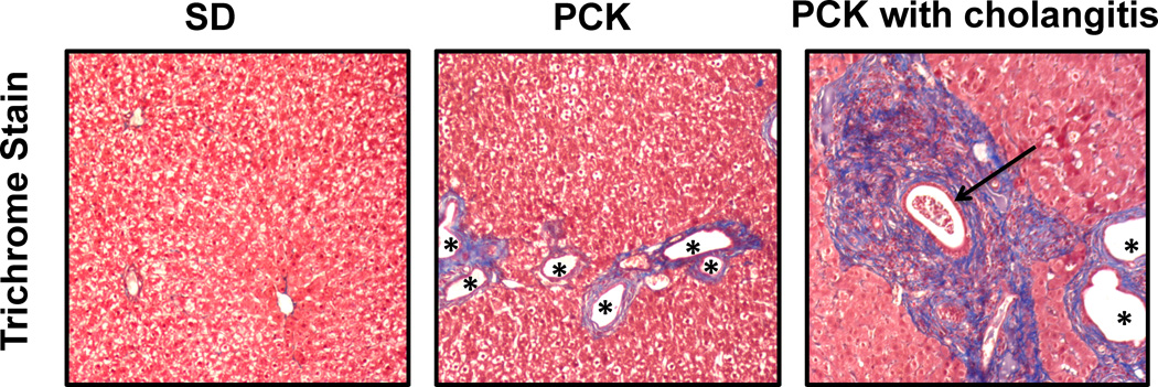
Figure 3.
Photomicrographs (10× original magnification) of SD and PCK rat liver specimens stained with Masson’s Trichrome to assess periportal fibrosis (in blue) and biliary dilatation (asterisks *). Note the regions of bile duct proliferation and dilatation as well as increased fibrosis in the PCK rats. Periportal fibrosis is especially pronounced in the rat with cholangitis, in which inflammatory cells are evident within a dilated bile duct (arrow).

