Figure 1.
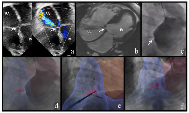
Image Fusion Guided Device Closure of Left Ventricle to Right Atrium Shunt. A. Pre-intervention trans-thoracic echocardiogram, 4-chamber view in color compare mode showing left ventricle to right atrium (LV-RA) shunt (white arrow). B. Pre-intervention steady-state free precession cardiac MRI (Siemens 1.5 Tesla) with noncontrast angiographic sequences, assisted by respiratory compensation and EKG-based cardiac gating. The magnetic resonance image shows right heart enlargement and LV-RA shunt (white arrow). The defect diameter is 3 mm and velocity-encoded MRI Qp:Qs is 1.4:1. C. The baseline conventional left ventriculogram shows the LV-RA shunt (white arrow). D. XFM [x-ray fused with MRI] overlay baseline left ventriculogram (blue = right ventricle, light pink = left ventricle, dark pink = LV-RA shunt) highlights the intracardiac shunt. E. Following XFM guided wire crossing of the LV-RA defect, this panel shows XFM guided defect closure with a muscular VSD occluder. The device is deployed in the defect and still attached to the delivery cable. F. The post-intervention left ventriculogram with XFM overlay shows successful device closure of the LV-RA defect.

